Appearance
Chapter 1: Understanding Bone Density Scans
1
What's a Bone Density Scan (DEXA)?
Think of it like a special X-ray that measures how much mineral is packed into your bones.
Here's the Thing Most People Don't Know...
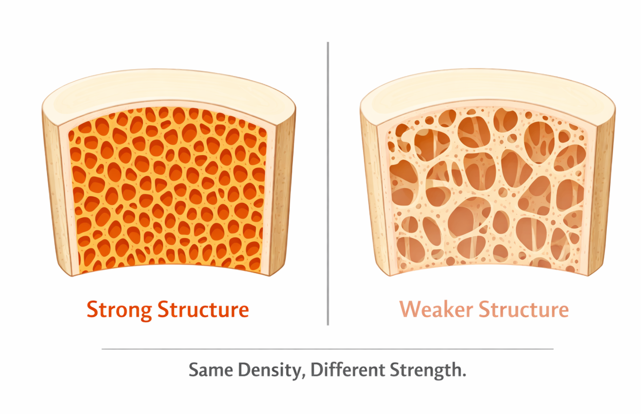
Bone density isn't the whole story. Two people can have the exact same density score, but one might have much stronger bones than the other. How? It's all about the internal structure.
Think of it like two buildings made with the same amount of concrete:
- Building A: The concrete is evenly spread throughout—strong and sturdy
- Building B: The concrete has big gaps and holes—same amount of material, but much weaker
Your bones are the same way. The scan measures how much mineral is there, but it can't see:
- How well the internal scaffolding is connected
- How thick or thin the supporting struts are
- Whether there are tiny cracks from daily wear and tear
- The quality of the collagen (the flexible stuff that keeps bones from snapping)
The Surprise Factor
This is why some people with "normal" scores still break bones, while others with low scores never do. Density is important, but it's only part of the picture.
Making Sense of Your Results
Your scan results come with two scores that compare you to other people:
| Score | What It Means |
|---|---|
| T-score | How you compare to a healthy 30-year-old |
| Z-score | How you compare to people your own age |
What the T-score numbers mean:
- -1 or higher: You're in great shape!
- -1 to -2.5: Your bones are a bit thinner than ideal (called osteopenia)
- -2.5 or lower: Osteoporosis territory—time to take action
The Petite Woman Paradox
Here's something that affects many smaller-framed people, especially women: DXA scans can make petite bones look worse than they really are.
Why? Because DXA measures areal density (grams per square centimeter)—it's looking at a 2D projection of a 3D bone. Because of the math involved, larger bones naturally produce a higher BMD score, even if the tissue density inside is exactly the same.
What this means in practice:
- A petite woman might show a low T-score
- But her actual bone tissue could be perfectly healthy (normal volumetric density)
- The "low" reading is partly an artifact of her smaller bone size
The Real Story
If you measured the same woman's bones with a 3D scan (QCT), you might find her volumetric bone density is completely normal. The DXA score looks low simply because her bones are smaller.
But here's the important catch: While the DXA score might be somewhat artifactual, the increased fracture risk from having smaller bones is real. Bone strength depends heavily on geometry—specifically diameter. A smaller bone, even with perfectly healthy tissue, is mechanically weaker than a larger bone.
Use the interactive tool below to see this in action—notice how dramatically bone diameter affects strength:
Cortical Bone Comparator
Visualize the impact of geometry on Bone Mineral Density (BMD) and Bending Strength.
Reference
Diameter: 25mmThickness: 5mm
Ref BMD100%
Ref Strength100%
Modified
BMD0.0%Cross-Sectional Mass
Strength (Bending)0.0%Moment of Inertia
25.0mm
10mm45mm
5.0mm
1mm20mm
Did you know? Moving mass away from the center (increasing diameter) increases bending strength exponentially (r⁴), much more than just adding thickness.
So petite women face a double challenge:
- Their DXA scores may look worse than their bone tissue quality deserves
- But their smaller bone geometry genuinely does put them at higher fracture risk
This is why doctors should interpret BMD results in context of body size, and why petite individuals may benefit from earlier attention to bone health—not because their bone tissue is necessarily unhealthy, but because their geometry offers less margin for error.
Beyond the Basic Scan
Good news: there are newer technologies that can look deeper:
Trabecular Bone Score (TBS): Uses your existing scan to analyze the texture pattern—kind of like looking at the weave of a fabric instead of just its weight.
REMS (Radiofrequency Echographic Multi Spectrometry): A newer ultrasound-based technology that measures both bone density AND bone quality—without radiation. It analyzes raw ultrasound signals from your spine and hip to assess the internal microarchitecture, not just mineral content. Studies show ~89% agreement with DXA for diagnosis, plus it provides a "Fragility Score" that predicts fracture risk independent of BMD. Because there's no radiation, it's safe for pregnant women, children, and frequent monitoring. The downsides: REMS is not widely available yet, often isn't covered by insurance, and isn't cheap. Also, while the technology looks promising on paper, some people report having good REMS scores yet still experiencing multiple fractures—a reminder that no scan perfectly predicts real-world bone strength. Important: never compare a REMS score to a DXA score—they measure differently and the numbers aren't interchangeable.
Special CT Scans (HR-pQCT): Can actually show individual bone structures in your wrist or ankle. These are available at special university clinics.
What About Fracture Risk?
Doctors often use a tool called FRAX that combines your bone density with other factors to estimate your chance of breaking a bone in the next 10 years. It considers things like:
- Your age and weight
- Whether you've broken bones before
- If your parents broke a hip
- Smoking and drinking habits
- Certain medications and conditions
The Bottom Line
Your bone density score is useful, but don't treat it as the final word on your bone health.
Here's what to remember:
- DXA scans measure mineral content—think of it as weighing your bones
- Two bones with identical scores can have very different strength
- If you've broken bones despite "normal" numbers, something else might be going on
- Your doctor should look at the full picture, not just one number
Up next, we'll explore what bones are actually made of—because understanding the basics helps everything else make sense!